Opportunities with radiotherapy
von Dr. Alena Soukup & Team // 24. January 2025
A tumor in the brain? Yes, our dogs can also get brain tumors. Brain tumors in dogs are not that rare, accounting for about 2-4.5% of the dog population. Theoretically, they can occur at any age. Since these tumors cannot be seen from the outside, they can only be detected by clinical signs and abnormalities. It is therefore all the more important to interpret clinical signs correctly and have them clarified by a veterinarian in good time.
Brain tumors in the strict sense originate directly from the nerve tissue of the brain. In a broader sense, this also includes all tumors within the cranium as well as the spinal cord and spinal canal.
Brain tumors in the strict sense are all tumors that originate directly from brain tissue. In a broader sense, all masses within the cranium that arise from uncontrolled cell growth are often referred to as brain tumors. In principle, such a tumor can originate from cells of all tissues and can exhibit varying degrees of aggressiveness and prognosis depending on the tissue of origin.
Tumors that originate from the meninges are referred to as meningiomas. These tumors are the most common tumors in the brain of our dogs and are formally considered benign. However, as they grow infiltratively, displace brain tissue and can exert pressure, these brain tumors are often very problematic in the broader sense.
When cells of the cranial supporting tissue degenerate and grow uncontrollably, astrocytomas or gliomas develop, which originate from the so-called glial cells. They are the second most common brain tumors in dogs.
The pituitary gland (hypophysis) can also develop tumors. These are divided into benign pituitary adenomas and malignant pituitary carcinomas and are also relatively common.
Other tumors such as histiocytic sarcomas, lymphomas and plasmacytomas are also possible in the brain.
Whether such a tumor is benign or malignant cannot be determined in veterinary medicine using imaging techniques alone. In general, a distinction is made between intra-axial masses, when the tumors originate directly from brain tissue, and extra-axial masses, when they do not originate from brain cells.
Brain tumors cannot be seen from the outside, so the suspicion always arises from clinical abnormalities. It is therefore important to clarify any neurological changes. Dogs with brain tumors are often presented with the following symptoms:
However, all symptoms can also have a different cause, so that self-diagnosis at home is not possible. A reliable diagnosis or the exclusion of a central nervous system problem using the correct examination method is particularly important for all these symptoms. The differences in the clinical symptoms depend primarily on the localization.
Tumors in the area of the cerebrum typically cause epileptic seizures. A tumor in the brain stem or cerebellum can be accompanied by gait disorders and uncoordinated movements. A pituitary adenoma can be active, in which case patients show typical symptoms of a disturbed hormone balance, e.g. hyperadrenocorticism, also known as Cushing's syndrome. Typical symptoms are increased drinking, combined with increased urination, muscle breakdown and poor coat quality. However, the adenoma can also be inactive. In this case, patients often present with fatigue (apathy), reduced appetite or, in advanced stages, blindness.
The neurological examination plays an important role in narrowing down the diagnosis. If the suspicion of a brain tumor is confirmed, imaging procedures are the method of choice for further diagnostics. With magnetic resonance imaging (MRI), the brain tissue can be visualized with very good contrast. The tumor itself is clearly visible and the displacement of the brain tissue due to a displacement effect of the mass can also be assessed. An additional swelling, known as edema, can often be seen around the brain tumor.
Alternatively, computer tomography (CT) can also be used if MRI is not available. However, the contrast is significantly poorer here and the diagnosis and differentiation is much more difficult in some cases. As with CT, a short anesthetic is also necessary for MRI in order to position the animal and to be able to carry out the examinations in high quality. In brain tumor patients, especially in animals that have already suffered a seizure, an increase in intracranial pressure must be avoided. Stress and excessive strain should be avoided. Ideally, the head should be slightly elevated even when lying down.
A definitive diagnosis by taking a sample (biopsy) is rarely carried out in veterinary medicine today, but sometimes the puncture of cerebrospinal fluid (cerebrospinal fluid) is helpful.
Magnetic resonance imaging is the imaging method of choice when examining the brain. This dog suffers from a brain tumor (right of the median). A clear shift of the midline shows the effects of this mass.
After a thorough neurological examination, modern imaging quickly and gently provides clarity about the extent of the changes. MRI is the imaging method of choice for brain tumors due to its high contrast. However, tumors can also often be very clearly delineated with CT. The image shows a tumor in the brain of a 12-year-old American Foxhound in a CT scan.
Around half of patients are diagnosed with a benign brain tumor, such as a meningioma or an adenoma of the appendage gland. Unfortunately, meningiomas can also show infiltrative growth in many cases and tend to come back after surgery, i.e. recur. Metastases are generally rather rare in brain tumors. The staging of these tumors nevertheless includes a chest X-ray and, if necessary, an abdominal ultrasound or a whole-body computed tomography scan. A brain tumor is always a problematic disease, as it compresses the brain tissue and, if the patient remains untreated, usually leads to death after a few months.
Depending on their location, brain tumors can be operated on, but it should not be forgotten that the majority of brain tumors show infiltrative growth. This infiltration is not treated by surgery, so in many cases the tumor cannot be completely removed. In these cases, it is a matter of time before the remaining cells form a new mass. In addition, operations on the brain are very demanding and associated with a high risk of complications. In contrast, radiotherapy can nowadays be carried out very well and safely in these localizations. The infiltrative part of the tumor is also treated, patients show a rapid clinical improvement in most cases and the published radiotherapy protocols rarely have significant side effects. At Equinox Healthcare, brain tumors are irradiated with 20 sessions or alternatively with 10 sessions. The radiotherapy takes place daily over four or two weeks.
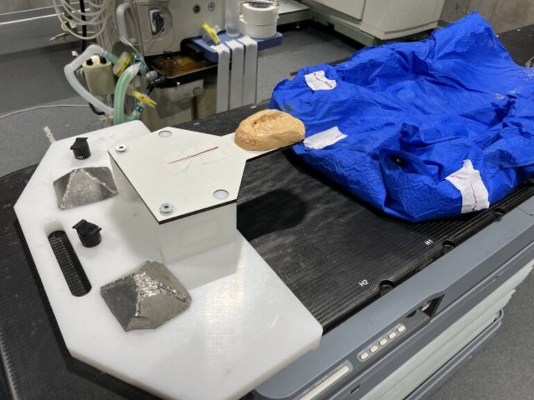
In order for a brain tumor to be irradiated, a computer tomography scan is required as a planning CT. A special positioning aid with a bite block, a dental impression and a vacuum mattress is produced. The sectional images are used to create an individual radiation plan in a special computer-based planning system. This procedure makes it possible to irradiate the tumor with a high dose while sparing the surrounding normal tissue.
For the accurate and precise irradiation of brain tumors, we use individual positioning aids such as vacuum mattresses, bite impressions and bite wedges. Plastic masks are only used at EQUINOX HEALTHCARE in exceptional cases, as they rarely improve accuracy.
There is no clear racial or gender predisposition for a brain tumor. In short-nosed, so-called brachycephalic breeds, intra-axial tumors occur more frequently than in other dog breeds.
The earlier a brain tumor is diagnosed, the better the chances of long-term tumor control. With radiotherapy, an average tumor control of two years with a very good quality of life is possible. The tumor does not necessarily have to disappear. A reduction or stabilization of the disease often leads to the disappearance of symptoms.